MIRI Detector Subarrays
MIRI imaging, coronagraphic imaging, and low-resolution spectroscopy utilize a pre-defined set of subarrays for different observing strategies, each with its own advantages and recommended uses.
On this page
See Also: Understanding Exposure Times
MIRI’s detector arrays have the ability to read out partial frames through the manipulation of clocking patterns. Subarray readouts reduce the frame time to less than the nominal 2.775 s per full frame. The portion of the full array that forms the subarray is read out and stored while the remaining parts of the array are reset.
There are 15 different subarrays available for imaging, coronagraphic imaging, and low-resolution spectroscopy (LRS): the FULL array, 4 separate subarrays for coronagraphs, 1 for high background, 5 for bright objects, 1 for LRS slit, and 3 for LRS slitless. This includes new subarrays for Cycle 6: 2 for bright objects, a new dedicated subarray for LRS slit, and 2 for LRS slitless. No subarrays are available for the medium-resolution spectrometer (MRS) or wide field slitless spectroscopy (WFSS).
Figure 1. Subarray locations for the MIRI imager as viewed from the telescope looking down onto the detector
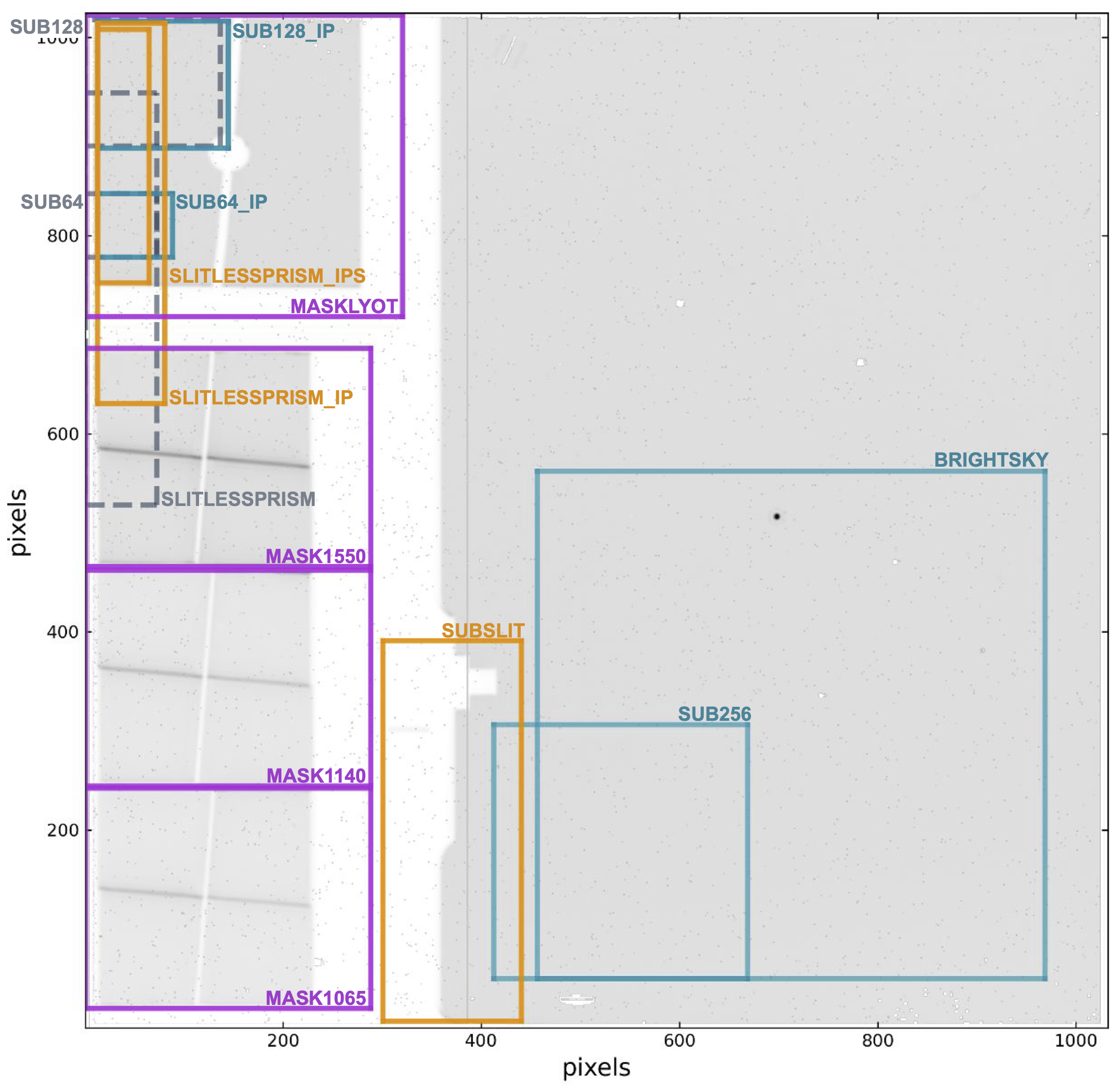
Click on the figure for a larger view.
Subarray locations on the imager detector for imaging (teal), LRS (gold), and coronagraphy (purple). All illustrated subarrays are available in Cycle 6. Subarrays in dashed grey lines will be retired in future cycles.
Subarray readout
See also: MIRI Detector Readout Overview, MIRI Detector Readout FASTR1, MIRI Detector Readout SLOWR1
Subarray coordinates cannot be randomly accessed so the row and column shift registers must step from the origin (1,1) to the starting subarray corner before proceeding. In other words, at the beginning of each frame read, the first two rows of the full array are accessed briefly and reset, then the 2nd pair, etc., until the subarray is reached.
In the first row of the subarray, pixels on the left that are not part of the subarray are clocked through (and not digitized), then the pixels that are part of the subarray are read. Pixels to the right of the subarray are ignored by resetting the column shift register to 0 immediately after the last subarray pixel. This pattern is repeated through all the rows contained within the subarray. The rows after the subarray are stepped through quickly and reset as were the rows before the subarray. It follows that subarrays are slower the farther they are from the left edge of the array.
This readout scheme drove the orientation of the imager array, since it is advantageous to have the fastest subarrays located within the coronagraph. It also means that it would normally take exactly the same amount of time to read a 256 × 256 subarray starting at (257,257) as it does to read a 512 × 256 subarray starting at (1,257); the only difference is the amount of data passed to the solid state recorders and the ground. However, for more efficient subarray operation, a "burst mode" clocks through the left columns at 5 times the normal speed. In the first row of the region of interest (ROI), pixels on the left that are not part of the ROI are clocked through (and not digitized), after which the pixels that are part of the ROI are read. Pixels to the right of the ROI are ignored by resetting the column shift register to zero (recall the special shift register definition) immediately after the last ROI pixel. This pattern is repeated through all the rows contained within the ROI. The rows after the ROI are stepped through quickly and reset as are the rows before the ROI.
Other issues that determined the subarray location included: the ability to utilize reference pixels, minimizing dead time from the clocking to access the first pixel, bad pixels on the array, and the best imaging location for the optics.
One known issue in the readout is the introduction of electromagnetic interference (EMI) into the rate images for some subarrays. This has to do with a characteristic noise frequency, usually 390 Hz, being out of phase with the time between pixel reads. EMI at 390 Hz affected two imaging subarrays (SUB128 and SUB64) and the LRS SLITLESSPRISM subarray. As of Cycle 6, new, re-shaped versions of these three subarrays are available in order to keep the 390 Hz noise in-phase with the pixel read times (see Brandt et al. 2025). The new subarrays have the suffix IP for "in-phase" and are named SUB128_IP, SUB64_IP, SLITLESSPRISM_IP, and SLITLESSPRISM_IPS.
Imaging
See also: MIRI Imaging, MIRI Imaging Template Parameters
Words in bold are GUI menus/
panels or data software packages;
bold italics are buttons in GUI
tools or package parameters.
Table 1. MIRI imaging subarray characteristics
| Subarray | Size in pixels (rows × columns) | First row corner | First column corner | Usable size | Group time† |
|---|---|---|---|---|---|
FULL | 1024 × 1032 | 1 | 1 | 74" × 113" | 2.775 s |
| BRIGHTSKY | 512 × 512 | 51 | 457 | 56.3" × 56.3" | 0.865 s |
| SUB256 | 256 × 256 | 51 | 413 | 28.2" × 28.2" | 0.300 s |
| SUB128 | 128 × 136 | 891 | 1 | 14.1" × 14.1" | 0.119 s |
| SUB128_IP | 128 × 132 | 889 | 13 | 14.1" × 14.1" | 0.120 s |
| SUB64 | 64 × 72 | 779 | 1 | 7" × 7" | 0.085 s |
| SUB64_IP | 64 × 76 | 779 | 13 | 7" × 7" | 0.087 s |
† Group times are calculated for FASTR1 mode only. SLOWR1 and FASTGRPAVG8 readout will only be available for the FULL array.
Coronagraphic imaging
See also: MIRI Coronagraphic Imaging, MIRI Coronagraphic Imaging Template Parameters
In coronagraphic imaging, each subarray is tied to a specific filter. The subarray choice is therefore hidden from the user in the JWST Astronomer's Proposal Tool (APT).
Table 2. MIRI coronagraphic imaging subarray characteristics
| Subarray | Filter | Size in pixels | First row corner | First column corner | Size‡ | Group time† |
|---|---|---|---|---|---|---|
| MASK1065 | F1065C | 224 × 288 | 21 | 1 | 24" × 24" | 0.240 s |
| MASK1140 | F1140C | 224 × 288 | 243 | 1 | 24" × 24" | 0.240 s |
| MASK1550 | F1550C | 224 × 288 | 463 | 1 | 24" × 24" | 0.240 s |
| MASKLYOT | F2300C | 304 × 320 | 719 | 1 | 30" × 30" | 0.324 s |
‡ This angular size is the illuminated FOV. The subarray size listed in pixels is the true size of the array (in pixels), which defines the exposure time per frame.
† Group times are calculated for FASTR1 mode only. SLOWR1 and FASTGRPAVG8 readout will only be available for the FULL array.
Wide field slitless spectroscopy
See also: MIRI Wide Field Slitless Spectroscopy
The wide field slitless spectroscopy (WFSS) mode uses the same FULL array as the imager.
Low-resolution spectroscopy
See also: MIRI Low Resolution Spectroscopy, MIRI Low Resolution Spectroscopy Template Parameters
The size of the SLITLESSPRISM subarray is determined by the number of pixels needed to cover the 5–14 μm low-resolution spectrometer spectrum in the dispersion direction and provide adequate sky observations for background subtraction in the spatial direction.
Starting in Cycle 6 there are three new subarray options for the LRS. The first is SLITLESSPRISM_IP, which is a redesigned version of SLITLESSPRISM that is unaffected by 390 Hz noise. SLITLESSPRISM_IP is smaller than SLITLESSPRISM and is located closer to the upper left corner of the imager detector; these two changes make for a slightly faster readout.
The second is SLITLESSPRISM_IPS, which is similar to SLITLESSPRISM_IP, with the "S" standing for "short" as this is a shorter, faster subarray designed to increase the number of groups per integration before saturation for the brightest sources. SLITLESSPRISM_IPS accomplishes a faster read time by sacrificing background in the cross-dispersion direction and wavelength coverage at the red end.
The third new subarray is for the LRS Slit. Prior to Cycle 6, observations in the LRS Slit had to be read out in FULL frame, which affected observations of bright targets. The new SUBSLIT subarray is smaller and is designed specifically for bright targets. Note that because the SUBSLIT subarray is far from the detector edge where readout occurs, it is not as fast as SLITLESSPRISM, SLITLESSPRISM_IP, or SLITLESSPRISM_IPS, but is still significantly faster than the FULL frame readout.
Table 3. MIRI low-resolution spectroscopy imaging subarray characteristics
| Subarray | Size in pixels | First row corner | First column corner | Group time† | Notes |
|---|---|---|---|---|---|
| FULL | 1024 × 1032 | 1 | 1 | 2.775 s | Slit spectrum |
| SUBSLIT | 384 × 140 | 8 | 301 | 0.279 s | Slit spectrum |
| SLITLESSPRISM | 416 × 72 | 529 | 1 | 0.159 s | Slitless spectrum |
| SLITLESSPRISM_IP | 384 × 68 | 631 | 13 | 0.156 s | Slitless spectrum |
| SLITLESSPRISM_IPS | 256 × 52 | 753 | 13 | 0.118 s | Slitless spectrum |
† Group times are calculated for FASTR1 mode only. SLOWR1 and FASTGRPAVG8 readout will only be available for the FULL array.
References
Brandt, T. D., Bergeron, E., Clarke, M. 2025, JWST-STScI-009091, SM-12
Electromagnetic Interference (EMI) in MIRI: Properties and Mitigation