MIRI Imaging
The MIRI imager offers 9 broadband filters covering wavelengths from 5.6 to 25.5 μm over an unobstructed 74" × 113" field of view, and a detector plate scale of 0.11 "/pixel (Bouchet et al. 2015). The MIRI imaging mode also supports the use of detector subarrays for bright targets, as well as a variety of dither patterns that could improve sampling at the shortest wavelengths, remove detector artifacts and cosmic ray hits, and faciliatate self-calibration. The Astronomer's Proposal Tool (APT) can be used to design mosaic observations to image larger fields.
On this page
Basic performance
See also: MIRI Performance, MIRI Sensitivity, MIRI Bright Source Limits
Imaging with MIRI is diffraction limited in all filters, with Strehl ratios in excess of 90%, although the detector plate scale of 0.11 "/pixel slightly undersamples the PSF in the F560W band.
MIRI imaging sensitivity is background limited in all the imaging bands (unless one takes short integrations): astronomical background limited at wavelengths <15 μm and telescope background (primary mirror and sunshield) limited at wavelengths >15 μm.
Observers will be able to specify settings for 4 primary MIRI imaging parameters: (1) filters, (2) dither pattern, (3) choice of subarray, and (4) detector read out modes and exposure time (via the number of frames and integrations).
Imaging field of view
The main MIRI imaging field of view (FOV) is 112.6" by 73.5" and is shown in Figure 1. In that figure, the grey regions on the left show the Lyot and the 4QPM FOVs, and have data in every imaging exposure. However, the 4QPM FOVs do not have valid data when the imaging filters are used, because these filters include additional optical elements that require special calibrations (i.e., separate calibrations designed specifically for the 4QPMs). Because the Lyot coronagraph has no additional optics, its FOV does provide valid, calibrated data except where the Lyot occulting spot and support structure prevent light from reaching the detector. The valid data regions for MIRI imaging exposures are shown in Figure 2.
Imaging filters
See also: MIRI Filters and Dispersers
Words in bold are GUI menus/
panels or data software packages;
bold italics are buttons in GUI
tools or package parameters.
Table 1. MIRI filter properties
Filter | λ0 | Δλ | FWHM* | Comment |
|---|---|---|---|---|
F560W | 5.6 | 1.00 | 0.207 | Broadband Imaging |
F770W | 7.7 | 1.95 | 0.269 | PAH, broadband imaging |
F1000W | 10.0 | 1.80 | 0.328 | Silicate, broadband imaging |
F1130W | 11.3 | 0.73 | 0.375 | PAH, broadband imaging |
F1280W | 12.8 | 2.47 | 0.420 | Broadband imaging |
F1500W | 15.0 | 2.92 | 0.488 | Broadband imaging |
F1800W | 18.0 | 2.95 | 0.591 | Silicate, broadband imaging |
F2100W | 21.0 | 4.58 | 0.674 | Broadband imaging |
F2550W | 25.5 | 3.67 | 0.803 | Broadband imaging |
* FWHM refers to the PSF
Figure 3. MIRI imaging filter bandpasses
Dithering performance
See also: MIRI Imaging Dithering, MIRI Dithering
MIRI operations offers several options for imaging dithers. There are multiple reasons for an observer to use dithers, some of which are unique to MIRI imaging.
Dithering allows for the removal of bad pixels and for improving the resolution of undersampled images. For MIRI imaging, only the F560W band produces undersampled images of point sources.
Dithering by a distance larger than a few times the PSF width on a timescale of a few minutes is necessary to self-calibrate detector gain variations and drifts since detector drifts grow larger with increasing signal.
At longer wavelengths, when the telescope background dominates the noise, dithering is needed to track temporal variations in the telescope background.
Multiple dither patterns are available to support different science strategies (e.g., deep imaging, snapshots, improved PSF sampling) and different target morphologies (e.g., point, compact and extended sources). They're also available for use with predefined detector subarrays.
As with the other near-infrared instruments, MIRI dither specifications can be conceptually separated into large- and small-scale dithers. Large-scale dithers are intended to handle self-calibration, large scale gain variations, and are useful to mitigate persistence and impact of cosmic rays. Since there is only one imaging MIRI detector, dithers are not required to cover gaps, as is the case of NIRCam. Small-scale dithers are needed to improve image quality when the native plate scale undersamples the PSF. For MIRI, only the F560W PSF is undersampled. The F770W PSF is Nyquist sampled and all other filters lead to oversampled PSFs.
Subarrays
See also: MIRI Detector Subarrays
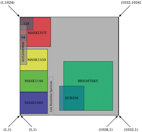
MIRI imaging supports a small pre-defined set of subarrays for imaging bright sources or bright backgrounds without saturating the detector. The MIRI imaging detector creates subarrays using a different scheme than the near-infrared HAWAII 2RG detectors that are used in other JWST instruments. In particular, frame time gets shorter as the subarray gets closer to one edge of the detector. For instance, coronagraphic subarrays are located on the fast side of the array, as are the smallest imaging subarrays, SUB128 and SUB64.
Subarray locations for the MIRI imager as viewed from the telescope looking down onto the detector. Imaging templates only provide access to the FULL, BRIGHTSKY, SUB256, SUB128, and SUB64 subarrays. The remaining subarrays are available for coronagraphic imaging (© Ressler et al. 2015).
| Subarray | Size (pixels) | Usable size (arcsec) | Frame time |
|---|---|---|---|
| FULL | 1024 × 1032 | 74" × 113" | 2.775 s |
| BRIGHTSKY | 512 × 512 | 56.3" × 56.3" | 0.865 s |
| SUB256 | 256 × 256 | 28.2" × 28.2" | 0.300 s |
| SUB128 | 128 × 136 | 14.1" × 14.1" | 0.119 s |
| SUB64 | 64 × 72 | 7" × 7" | 0.085 s |
Imager exposure specifications
See also: MIRI Detector Readout Overview, Understanding Exposure Times
MIRI imaging supports 2 different detector readout patterns:
References
Bouchet, P. et al. 2015, PASP, 127, 612
The Mid-Infrared Instrument for the James Webb Space Telescope, III: MIRIM, The MIRI Image
Ressler, M.E. et al. 2015, PASP, 127, 675
The Mid-Infrared Instrument for the James Webb Space Telescope, VIII: The MIRI Focal Plane Syste
Rieke, G. et al. 2015, PASP, 127, 584
The Mid-Infrared Instrument for the James Webb Space Telescope, I: Introductio


